Activation of 5-HT2A receptors upregulates the function of the neuronal K-Cl cotransporter KCC2
Résumé
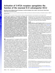
In healthy adults, activation of γ-aminobutyric acid (GABA) A and glycine receptors inhibits neurons as a result of low intracellular chloride concentration ([Cl-] i), which is maintained by the potassium chloride cotransporter KCC2. A reduction of KCC2 expression or function is implicated in the pathogenesis of several neurological disorders, including spasticity and chronic pain following spinal cord injury (SCI). Given the critical role of KCC2 in regulating the strength and robustness of inhibition, identifying tools that may increase KCC2 function and, hence, restore endogenous inhibition in pathological conditions is of particular importance. We show that activation of 5-hydroxytryptamine (5-HT) type 2A receptors to se-rotonin hyperpolarizes the reversal potential of inhibitory postsyn-aptic potentials (IPSPs), E IPSP , in spinal motoneurons, increases the cell membrane expression of KCC2 and both restores endogenous inhibition and reduces spasticity after SCI in rats. Up-regulation of KCC2 function by targeting 5-HT 2A receptors, therefore, has therapeutic potential in the treatment of neurological disorders involving altered chloride homeostasis. However, these receptors have been implicated in several psychiatric disorders, and their effects on pain processing are controversial, highlighting the need to further investigate the potential systemic effects of specific 5-HT 2A R ago-nists, such as (4-bromo-3,6-dimethoxybenzocyclobuten-1-yl)methyl-amine hydrobromide (TCB-2). T he neuron-specific K +-Cl-cotransporter KCC2 (encoded by the solute carrier family 12 member 5, Slc12a5) extrudes Cl-and is responsible for the low [Cl-] i in mature neurons (1-3), a prerequisite for hyperpolarizing inhibition mediated by GABA A receptors (GABA A Rs) and glycine receptors (GlyRs). The expression or the function of KCC2 is reduced in several neurological disorders (2, 4), and the resulting slight increase in [Cl-] i (depola-rizing shift of the chloride equilibrium potential, E Cl) dramatically compromises the inhibitory control of firing rate and excitatory inputs (5-7). Given the role of KCC2 in regulating the strength of inhibitory synaptic transmission, identifying tools that may increase KCC2 function and, hence, restore endogenous inhibition in pathological conditions is of particular importance. Spasticity is a disabling complication affecting individuals with spinal cord injury (SCI) and is characterized by a velocity-dependent increase in muscle tone resulting from hyperexcitable stretch reflexes, spasms, and hypersensitivity to normally innocuous sensory stimulations (8, 9). Down-regulation of KCC2 after SCI in rats is implicated in the development of spasticity (10) and chronic pain (11, 12). Notably, the expression of KCC2 in the motoneuron membrane is reduced, and, concomitantly, the density of cyto-plasmic clusters is higher, suggesting that the surface stability of the transporter is reduced in these pathological conditions (10). Mounting evidence indicates that phosphorylation of KCC2 in the C-terminal intracellular domain dynamically regulates its activity and surface expression (1). In particular, phosphorylation by protein kinase (PK)C, enhances KCC2 activity and reduces endo-cytosis (13). Interestingly, activation of 5-hydroxytryptamine type 2 receptors (5-HT 2 Rs) to serotonin stimulates PKC and strengthens the left-right alternation of motor bursts observed during loco-motion (14-16), which rely on reciprocal inhibition (17, 18). We hypothesized that 5-HT 2 R activity modulates KCC2 function and/ or expression. Our results indicate that the activation of the 5-HT 2A R subtype hyperpolarizes E IPSP via a PKC-dependent mechanism , increases KCC2 expression in the plasma membrane of motoneurons, and reduces SCI-induced spasticity. Results Negative Shift of E IPSP and Up-Regulation of KCC2. We first examined the effect of the 5-HT 2A/2B/2C R agonist (±)-2,5-dimethoxy-4-iodoamphetamine hydrochloride (DOI) (10 μM; Table S1) on E IPSP in control neonatal rats [postnatal day (P)5-P7]. DOI-hyperpolarized E IPSP within 10-20 min (Fig. 1 A and B). This effect was long-lasting (at least 2 h; Fig. 1B). E IPSP was significantly more hyperpolarized when motoneurons were recorded in the presence of DOI compared with control (8 mV; Fig. 1C, Left). There was a concomitant trend toward a depolarization of the resting membrane potential (V rest) by DOI (+2 mV; P > 0.05). As a result, the amplitude of hyperpolarizing IPSPs recorded at V rest increased significantly (Fig. 1C, Right). The next series of experiments was performed on animals that underwent a neonatal SCI. E IPSP was significantly more depolar-ized in those animals tested at P5-P7, compared with controls of the same age, as shown previously (19) (compare Fig. 1 C and D). Because of the increased sensitivity of neurons to 5-HT in those animals (15), DOI was tested at a lower concentration (1-1.5 μM) than in controls. DOI induced an ∼8-mV hyperpolarization of E IPSP (Fig. 1D, Left). As a result, E IPSP shifted from above to below V rest (Fig. 1D, Right). Another set of animals was treated chronically with DOI from P4 to P6-P7 [0.15 mg/kg, i.p. (15, 20) twice a day]. E IPSP was more hyperpolarized in those DOI-treated animals than in untreated transected animals (Fig. 1E). Values were similar to those measured in control animals. We performed subcellular fractionation of proteins from the lumbosacral spinal cord, followed by immunoblotting with a specific antibody against KCC2. The amount of KCC2 in the membrane fraction (KCC2Mb) was significantly increased after chronic DOI treatment, compared with NaCl-treated pups (Fig. 1F). There was a trend toward an increase in the amount of KCC2 in the cytosolic fraction. As a result, the ratio KCC2Mb/KCC2 cy-toplasm was nonsignificantly increased. We then analyzed the expression of KCC2 by immunohistochemistry. All of the analyses were performed on a homogeneous population of retrogradely labeled lumbar motoneurons [triceps surae (TS) muscles (ankle extensors); Fig. 1G]. GlyRs are colocalized with the anchoring protein gephyrin and can, therefore, be used to label the plasma Author contributions: R.B. and
Fichier principal
 Bos et al. - 2013 - Activation of 5-HT2A receptors upregulates the fun.pdf (12.56 Mo)
Télécharger le fichier
Bos et al. - 2013 - Activation of 5-HT2A receptors upregulates the fun.pdf (12.56 Mo)
Télécharger le fichier
Origine : Fichiers produits par l'(les) auteur(s)
Loading...

